


●Lieferzeit: ca. 3-5 Werktage **
Fast and reliable shipping
Need help? Check our
Service point
or
Contact us
| Item No.: | 23196-01 |
|---|---|
| Manufacturer item no.: | 4800 |
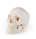
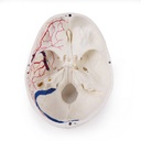
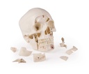
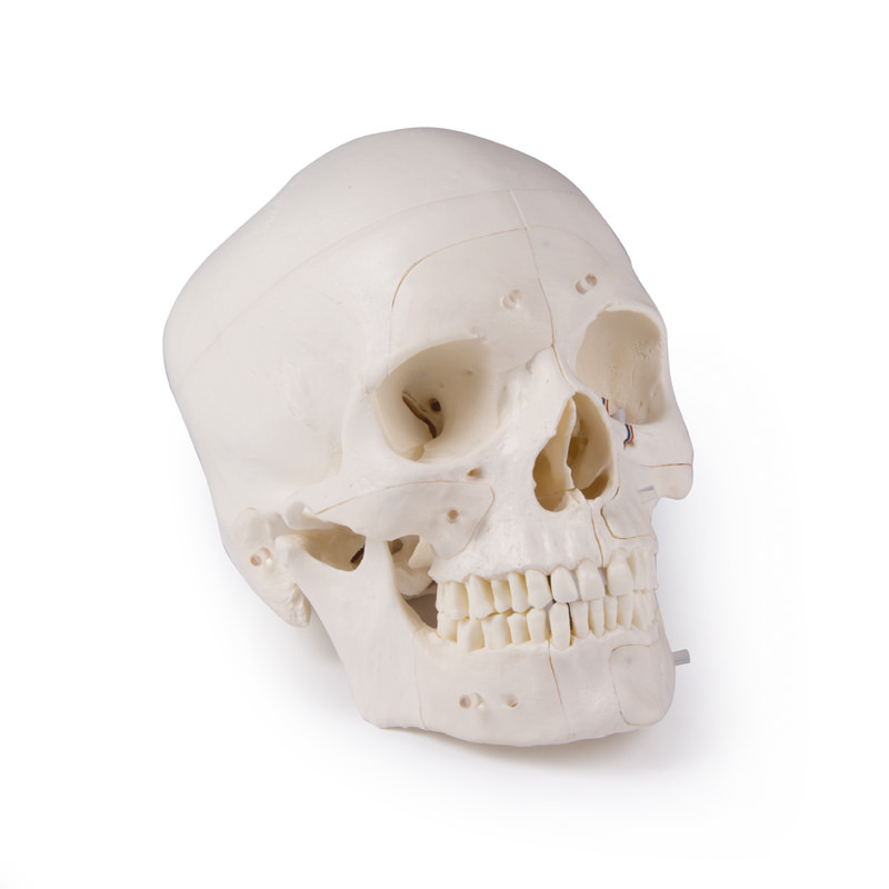
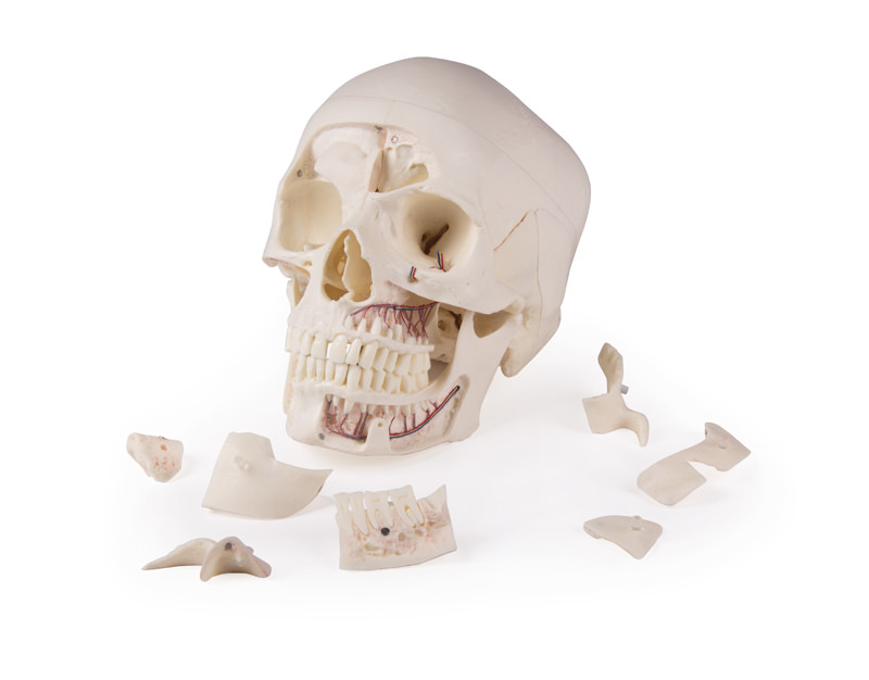
This skull model is a cast of a real human skull specimen and shows all anatomical structures with the highest level of detail. It was developed for students of anatomy, medicine, surgery, ENT, ophthalmology and dentistry. The skull is cut in a complex manner and assembled using metal and magnetic connections.
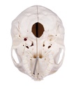
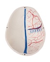
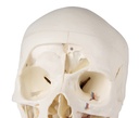
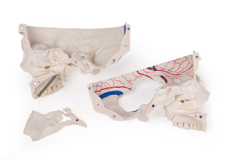
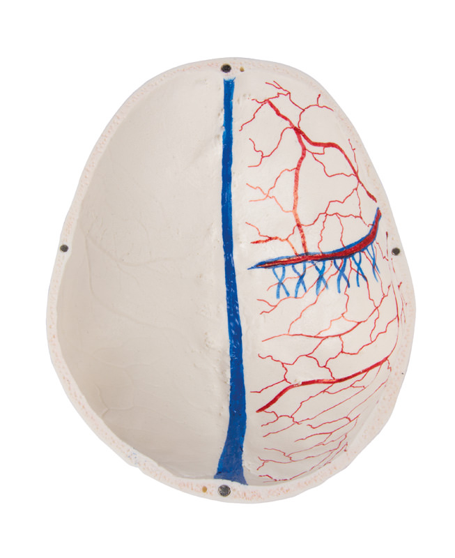
The calvaria is opened and removable, while leaving the temporal bones and their sutures intact. Bony impressions of the superior sagittal sinus, transverse sinus and sigmoid sinus, as well as the meningeal vessels, are painted. The skull base is sagittally sectioned so that on one side the cut passes through one cribriform plate and, at the same level, a further cut passes through the other cribriform plate of the ethmoid bone, leaving the crista galli and the perpendicular plate as well as the entire nasal septum intact. The structures of the anterior, middle and posterior cranial fossae are easily accessible. The nasal cavity, nasal conchae, septum, as well as the bony pharyngeal space and nasopharynx can be seen directly. The nasal septum can be removed from the surrounding bony structures. The frontal sinuses are prepared in one piece on one side; on the other side they are opened so that they are fully accessible. The relationship of this sinus to the nasal cavity is clearly visible; this is particularly valuable for ENT specialists.
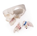
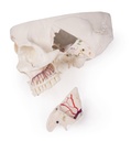
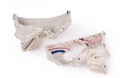
On one side of the skull, the temporal bone has been left in situ. The other temporal bone is removable from the skull. Part of the mastoid process and the squamous part of the temporal bone, together with the mastoid antrum, can be removed and provide a clear view of the inner ear. All three semicircular canals are visible, together with the course of the facial nerve, which runs posteriorly and then inferiorly and ultimately exits through the stylomastoid foramen. The removable temporal bone shows a complete external auditory canal. An almost vertical cut through the mastoid process and then further medially along the petrosquamous fissure divides the temporal bone and the position of the tympanic membrane can be seen. The carotid canal as well as the cochlea are opened and show the internal auditory canal, and the course of the facial nerve can be identified. The oval window, the semicircular canals and the opening of the tympanic cavity can be identified.
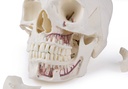
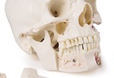
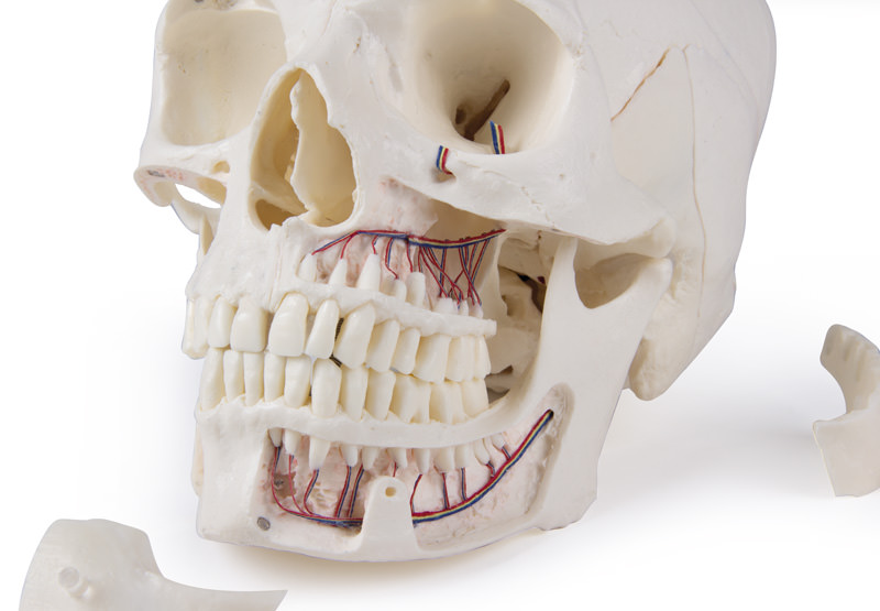
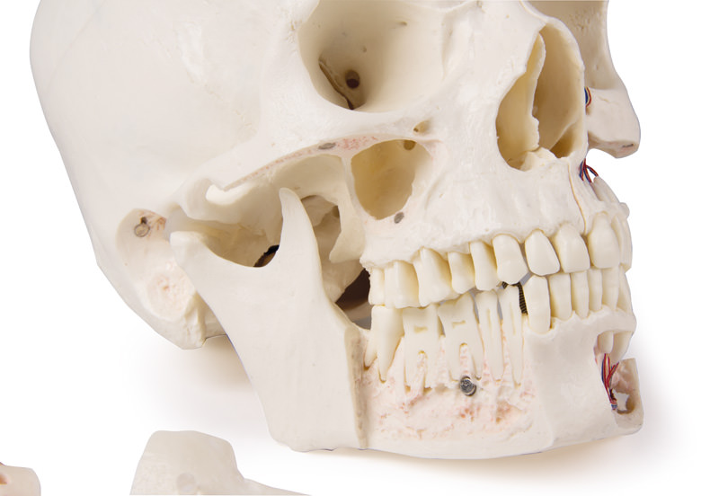
The maxilla and mandible show the structures of the dentition, the roots, the bony margin of the alveolar process, as well as dental nerves and vessels. The maxillary sinus can be opened by removing a flap. The teeth of the right mandibular flap are sectioned and show the internal structures of the teeth.
The calvaria is opened and removable, while leaving the temporal bones and their sutures intact. Bony impressions of the superior sagittal sinus, transverse sinus and sigmoid sinus, as well as the meningeal vessels, are painted. The skull base is sagittally sectioned so that on one side the cut passes through one cribriform plate and, at the same level, a further cut passes through the other cribriform plate of the ethmoid bone, leaving the crista galli and the perpendicular plate as well as the entire nasal septum intact. The structures of the anterior, middle and posterior cranial fossae are easily accessible. The nasal cavity, nasal conchae, septum, as well as the bony pharyngeal space and nasopharynx can be seen directly. The nasal septum can be removed from the surrounding bony structures. The frontal sinuses are prepared in one piece on one side; on the other side they are opened so that they are fully accessible. The relationship of this sinus to the nasal cavity is clearly visible; this is particularly valuable for ENT specialists.
On one side of the skull, the temporal bone has been left in situ. The other temporal bone is removable from the skull. Part of the mastoid process and the squamous part of the temporal bone, together with the mastoid antrum, can be removed and provide a clear view of the inner ear. All three semicircular canals are visible, together with the course of the facial nerve, which runs posteriorly and then inferiorly and ultimately exits through the stylomastoid foramen. The removable temporal bone shows a complete external auditory canal. An almost vertical cut through the mastoid process and then further medially along the petrosquamous fissure divides the temporal bone and the position of the tympanic membrane can be seen. The carotid canal as well as the cochlea are opened and show the internal auditory canal, and the course of the facial nerve can be identified. The oval window, the semicircular canals and the opening of the tympanic cavity can be identified.
The maxilla and mandible show the structures of the dentition, the roots, the bony margin of the alveolar process, as well as dental nerves and vessels. The maxillary sinus can be opened by removing a flap. The teeth of the right mandibular flap are sectioned and show the internal structures of the teeth.
Kontaktinformationen im Rahmen der Produktinformationsverordnung (GPSR)
Gesetzliche Anbietererkennung:
Erler-Zimmer GmbH & Co.KG
Hauptstrasse 27, 77886 Lauf, Deutschland
Mailkontakt: nicolas.kaltenbach@erler-zimmer.de
Achtung! Medizinisches Ausbildungsmaterial, kein Spielzeug. Nicht geeignet für Personen unter 14 Jahren.
Attention! Medical training material, not a toy. Not suitable for persons under 14 years of age.
Products from Erler-Zimmer
Erler-Zimmer offers high-quality, handcrafted models and medical teaching aids for human and veterinary medicine, as well as practice-oriented training materials used worldwide in the education, further training and continuing professional development of medical professionals.
Ihr dynamisches Snippet wird hier angezeigt ...
Diese Meldung wird angezeigt, weil Sie weder einen Filter noch eine Vorlage zur Verwendung bereitgestellt haben.